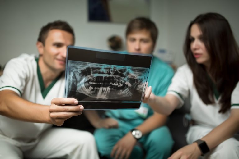
Thanks to technology, we have tools that can help us prevent, detect, and treat diseases. Doctors use diagnostic tools, such as scans and ultrasounds, to figure out what’s happening inside our bodies. There are different types of heart scans that physicians use to diagnose their patients. In London, there are private ultrasound scans that can help patients understand their condition.
Otherwise known as coronary calcium scoring, a heart scan can be described as an x-ray test for your heart. Physicians use this type of scan to check for calcium deposits in the arterial walls.
The calcium-containing plaque inside your heart’s arteries can build up over time and restrict blood flow to your heart muscles. Heart scans enable doctors to identify possible heart diseases before any signs or symptoms manifest. Here are some ways your physician can check your heart.
1. Chest X-rays
X-rays produce images of bones, tissues, and organs of the body. They utilise photons to cast shadows and show outlines of the human body.
A medical practitioner can use a chest x-ray to check for emphysema, lung cancer, pneumonia, and other severe conditions.
If you experience chest pains, a persistent cough, or shortness of breath, you need to get a chest x-ray. Chest x-rays are a quick and easy process that requires minimal preparation.
2. CT Scans

Computerized tomography scans use information from x-rays with 3D images of your body. The different types of CT scans used to check for problems with the heart include coronary CT angiograms (CTAs), calcium-score screening heart scans, and total body CT scans.
CTAs are non-invasive heart imaging tests that offer high-resolution 3D pictures of great vessels and the heart. This type of CT scans helps doctors determine if there’s an accumulation of calcium deposits of cholesterol in your coronary arteries.
Calcium-scoring screening heart scans are often used to detect coronary calcification from atherosclerosis – an ailment characterised by cholesterol, fats, and other substances building inside the arteries.
Total body CT scans (TBCTs) give physicians a detailed look inside the body. The scans analyse the heart, pelvis/abdomen, and the lungs. Using TBCT images, your doctor can detect aortic aneurysms (enlargement of the aorta).
3. Echocardiograms
An echocardiogram is also known as echocardiography, an echo, or a cardiac echo. It is a medical diagnostic tool described as an ultrasound of the heart.
Cardiac echos use high-frequency sound waves to produce images of the heart, which enable physicians to check the function and structure of your heart. If you’re experiencing breathlessness or chest pains, an echo would be an essential tool to help you ascertain the root cause of your health problems.
The different types of echocardiograms include stress, foetal, trans-thoracic, and trans-oesophageal echocardiograms.
- Stress echos can pick out heart problems such as heart valve dysfunction.
- Doctors use foetal echocardiograms to check the heart of a developing baby.
- Trans-thoracic echocardiography offers comprehensive information about the heart’s function and structure.
- Trans-oesophageal echocardiograms (TOE) require cardiac monitoring and sedation. The procedure helps doctors assess heart valves and check for blood clots in the heart chambers.
Medicine offers a myriad of solutions that can help you enjoy a healthy lifestyle. But people need to see their doctors at least once a year to make sure they are in the best of health, and to prevent an illness from becoming more serious.

